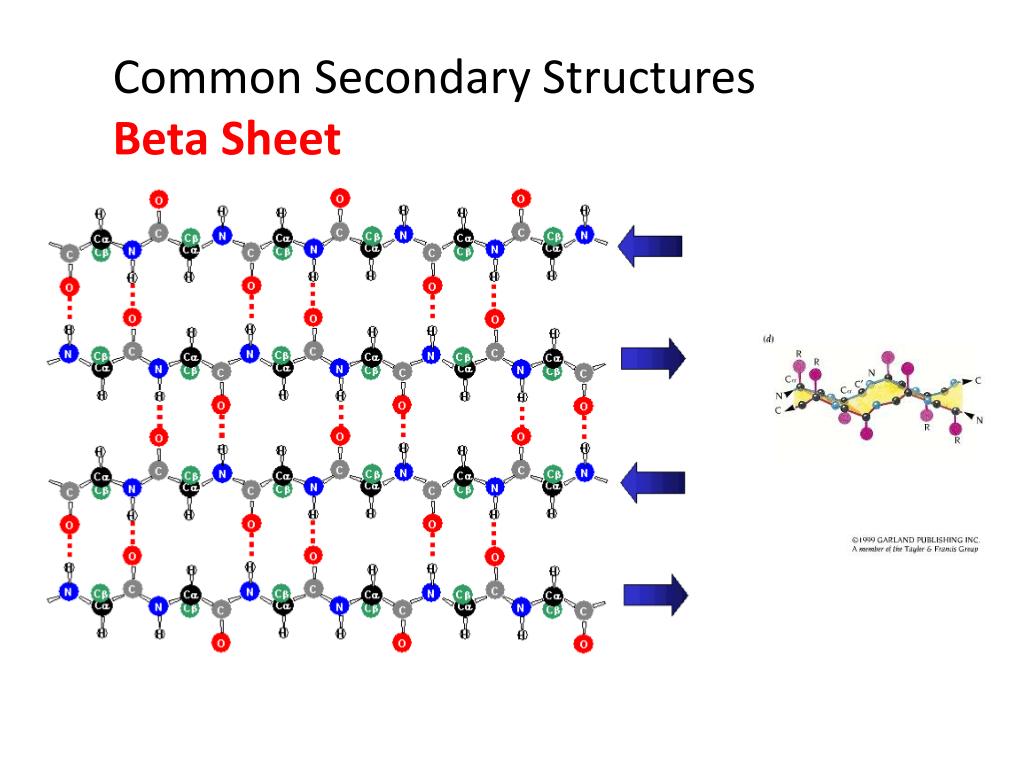
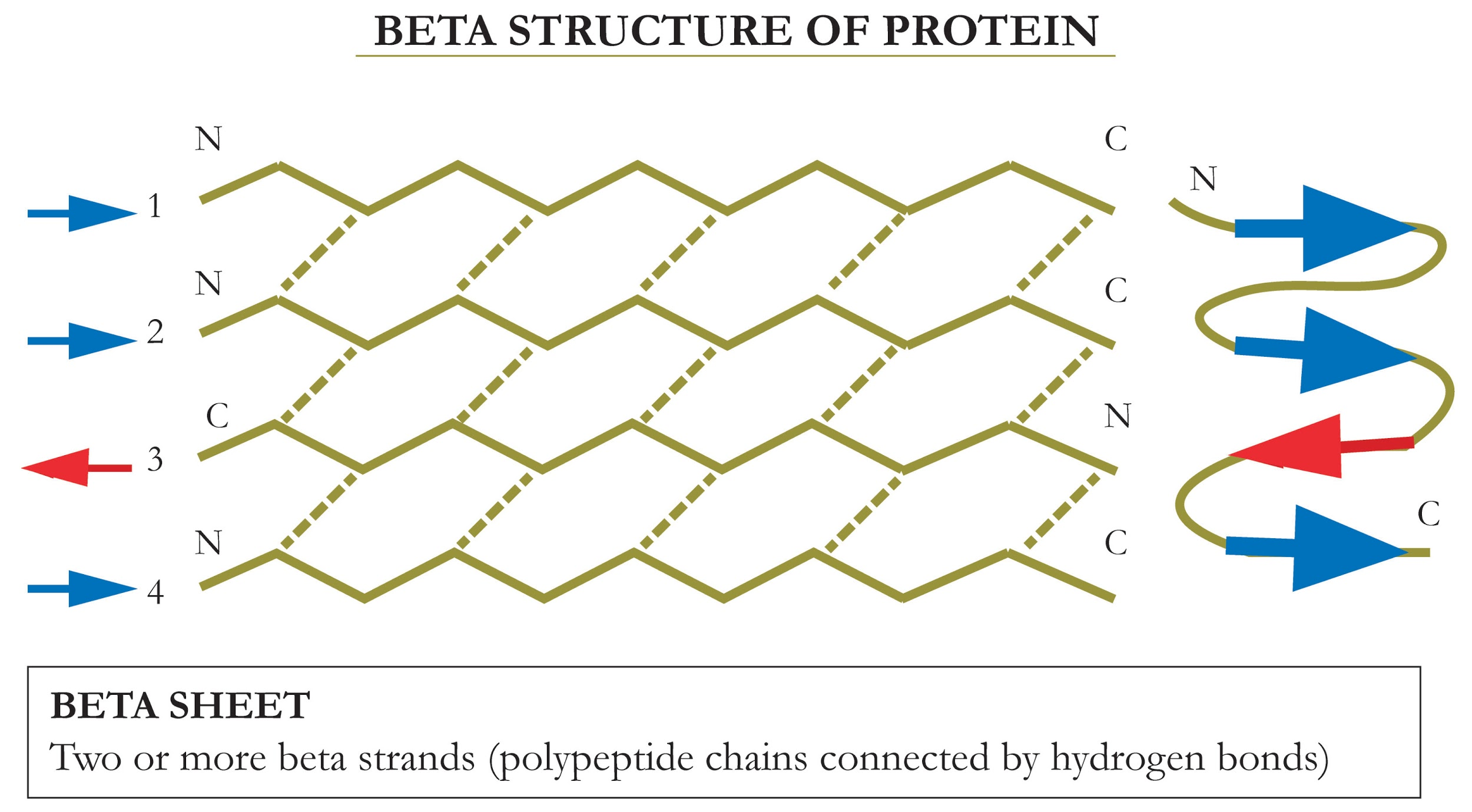
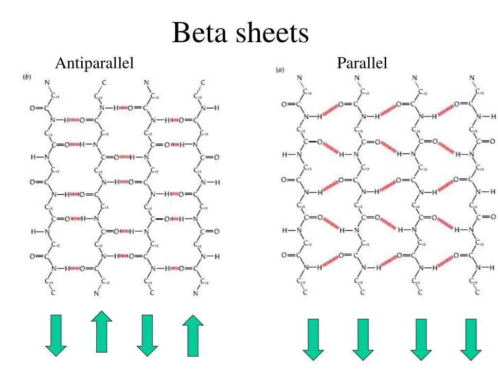
Beta Sheet Structure
Beta Sheet Structure - See examples of protein structures in stick and ribbon representations, and how they are related to the ramachandran plot and the 3d structure. Explore the four levels of protein structure, including secondary structure with α helix and β pleated sheet, and see examples and diagrams. Parallel sheets characteristically distribute hydrophobic side chains on both sides of the sheet, while antiparallel sheets are usually arranged with all the hydrophobic residues on one side. Web learn about beta sheet, a type of secondary structure formed from beta strands by hydrogen bonding. Web learn how proteins get their shape and function from the sequence and interactions of amino acids. Web the side chains in the beta sheet are normal to the plane of the sheet, extending out from the plane on alternating sides. Explore various aspects of beta sheet structure, formation, and applications in biochemistry and biotechnology.

Characterizing the αsheet Structure Daggett Research Group
PPT Protein Structure and Prediction PowerPoint Presentation, free
The beta pleated sheet structure of protein is due to(a)Formation of
PPT Introduction to Protein Structure PowerPoint Presentation, free

1. Secondary structure of protein, αhelix and βpleated sheet [118

Illustrated Glossary of Organic Chemistry Beta sheet, betapleated sheet

Chapter 2 Protein Structure Chemistry

Biochemistry Fundamentals Secondary Structure 2 The Beta Sheet

College. Science. Life Essential Cell Biology 3rd Ch 4 Protein

Secondary structures of keratin protein (beta pleated sheets and alpha
Web Learn How Proteins Get Their Shape And Function From The Sequence And Interactions Of Amino Acids.
Web learn about beta sheet, a type of secondary structure formed from beta strands by hydrogen bonding. Explore various aspects of beta sheet structure, formation, and applications in biochemistry and biotechnology. Explore the four levels of protein structure, including secondary structure with α helix and β pleated sheet, and see examples and diagrams. Parallel sheets characteristically distribute hydrophobic side chains on both sides of the sheet, while antiparallel sheets are usually arranged with all the hydrophobic residues on one side.
See Examples Of Protein Structures In Stick And Ribbon Representations, And How They Are Related To The Ramachandran Plot And The 3D Structure.
Web the side chains in the beta sheet are normal to the plane of the sheet, extending out from the plane on alternating sides.